You are here

User Stories

Haley Nation, a graduate student and anatomy teaching assistant to medical and physician assistant students, had her “a-ha” moment when she modeled and printed a CD replica of a section of the spinal cord:

Throughout my teaching, I have noticed students frequently struggle with understanding the anatomy of the sympathetic nervous system (and rightfully so!). This is a difficult dissection in the cadaver laboratory and therefore often not well appreciated. I thought that printing a simplified model would help students understand the potential neuronal pathways. I used TinkerCad to create the CD model and I found that even my understanding of the anatomy was enhanced.

In fact, this experience was so impactful for her learning, that a workshop for all anatomy teaching assistants is being planned in conjunction with Hector Lopez, MD Director of Human Structure and Integration.

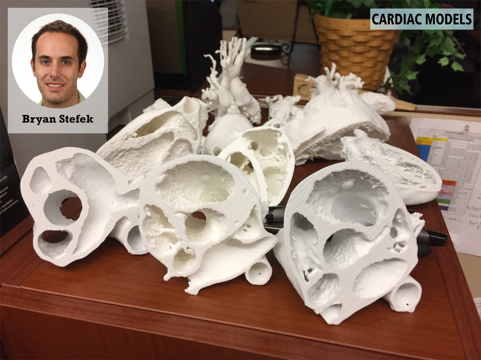
Bryan Stefek, MD a pediatric cardiology fellow, used technology in the library to create 3D-printed models of pediatric hearts from CT scans and MRI images.

I was interested in using this technology for didactic purposes; specifically teaching families, residents, fellows, and medical students about complex cardiac anatomy. To date, I have printed twelve 3D cardiac models both with normal and abnormal cardiac anatomy. I used Blender, a program which allows users to alter 4D models for eventual printing. I sliced 3D models to replicate images obtained during echocardiography.

Training on all these task was provided by the library. 

Find out more about 3D Printing at the Technology Sandbox.


Amy Ricords, BSN, MEd, Director of Nursing Education and Professional Development used the One Button Studio to record a training video for nursing staff:

I often repeat large conferences to reach all Nursing learners who work 24 hours a dayT 7 days a week. A particular speaker was unable to be present for all of the repeated sessions but her information was too valuable to skip. Instead of having her sit in front of a camera and lecture, we used the One Button Studio. One Button permitted the presentation to look so professional and much more engaging for the learner.

Patricia Silveyra, PhD, and her medical student partners were some of the first users of our One Button Studio to produce a training video for medical interpreting. They used the library’s video production suite to edit out mistakes, include closed captioning, and insert a still image of an exam room for a realistic background.

None of us had any experience using the software or the studio, but were able to film and ultimately produce a great product!
- Chavely Valdes-Sanchez, Medical Student Class of 2019

Aman Dhawan, MD, and Christopher Arena, MD produced a video which they submitted as supplementary material for a journal article on a specific surgical technique.

Dr. Arena stated:

One Button Studio and Ryan Klinger specifically were amazing in helping us to edit video and include voice over. Ryan spent a lot of time walking me through the steps and educating me on the software… and I would love to use it again in future projects.

Find out more about the One Button Studio.


Two clinical faculty used the large-scale display capability of our 3x3 video wall to enhance learning related to medical imaging.

Hector Lopez, MD used the video wall to show side-by-side comparisons of 3D and MRI brain images while teaching an outreach session for high school students interested in brain injury. On one side were MRI images from the library's web-based e-anatomy platform, IMAIOS e-anatomy. Dr. Lopez used the library’s Solstice app to wirelessly connect his iPad to the video wall, allowing him to manipulate the MRI images on his iPad while walking throughout the room interacting with students rather than being tied to the podium. Juxtaposed with these images were 3D slices of the brain from the library's Visible Body platform, so that students could visually compare the images.


Similarly, in his elective course for 4th year medical students on advanced musculoskeletal imaging, Cayce Onks, MD used the video wall while teaching students how to perform ultrasound imaging. Several ultrasound machines were set up in the room and the images from each displayed simultaneously on the video wall., which allowed learners to compare their technique with others in real
time.

I thought being able to integrate two types of technologies to enhance the teaching experience was great. I was able to run my power point with live musculoskeletal scanning being displayed on the other side of the screen. I have done similar presentations at regional conferences, but that required two projectors and two screens in a large conference room. This obviously was much better quality.

Find out more about the 3x3 video wall at the Technology Sandbox.


Brennan Ross, a physician assistant student who describes himself as “tech obsessed,” has introduced his student colleagues to some of the library’s virtual reality applications available in the Technology Innovation Sandbox using our VIVE headset.

"This makes so much more sense than learning anatomy from a picture. It’s even better than a screen where you can move it in a 3D plane. For example, there are two ways that you’re always being shown the spine in images, but you don’t know how they relate to each other and how they translate. Virtual reality and 3D printing help you see instantly how they relate.

Find out more about the Virtual Reality at the Technology Sandbox.