Haley Nation, a graduate student and anatomy teaching assistant to medical and physician assistant students, had her “a-ha” moment when she modeled and printed a CD replica of a section of the spinal cord:
Throughout my teaching, I have noticed students frequently struggle with understanding the anatomy of the sympathetic nervous system (and rightfully so!). This is a difficult dissection in the cadaver laboratory and therefore often not well appreciated. I thought that printing a simplified model would help students understand the potential neuronal pathways. I used TinkerCad to create the CD model and I found that even my understanding of the anatomy was enhanced.
In fact, this experience was so impactful for her learning, that a workshop for all anatomy teaching assistants is being planned in conjunction with Hector Lopez, MD Director of Human Structure and Integration.
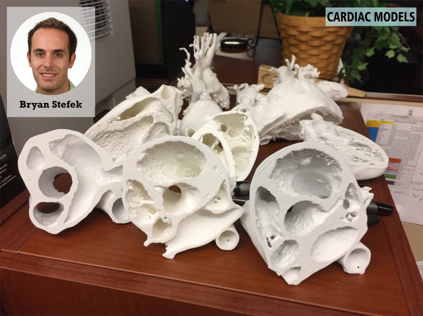
Bryan Stefek, MD a pediatric cardiology fellow, used technology in the library to create 3D-printed models of pediatric hearts from CT scans and MRI images.
I was interested in using this technology for didactic purposes; specifically teaching families, residents, fellows, and medical students about complex cardiac anatomy. To date, I have printed twelve 3D cardiac models both with normal and abnormal cardiac anatomy. I used Blender, a program which allows users to alter 4D models for eventual printing. I sliced 3D models to replicate images obtained during echocardiography.
Training on all these task was provided by the library.
Find out more about 3D Printing at the Technology Sandbox.
